
Read more about thyroid eye disease, including recent research and case studies from Endocrine Practice and AACE Clinical Case Reports.
Study: Assessing the Management of Thyroid Eye Disease-Related Strabismus

Thyroid eye disease (TED)-related strabismus warrants careful evaluation and management and comprises several surgical and non-surgical options to achieve an optimal outcome, according to a review published in the Journal of Current Ophthalmology.
Researchers combed PubMed, Google Scholar, and Ovid Medline for keywords, including TED-related strabismus; strabismus in thyroid-associated ophthalmopathy; Graves’ ophthalmopathy-related strabismus or squint; and restrictive strabismus. Two expert strabismus specialists selected and evaluated 115 articles published in the past 20 years, of which 98 were related to the topic of the review. The researchers categorized management of TED-related strabismus as surgical and non-surgical.
The results of the review showed that botulinum toxin A (BTA) is an effective method of non-surgical management of strabismus in active TED and residual deviation following surgery. The researchers observed that postoperative under-correction is more common in treating TED-related esotropia. They also listed lateral rectus resection and BTA as options to manage the problem.
Additionally, strabismus specialists recommended rectus muscle resection should be performed following maximum recession of restricted muscles, but this approach should be avoided on a restricted or enlarged muscle.
“Management of strabismus is challenging in TED. The following assessments should be considered, including ocular ductions, the amount of deviation, frequency doubling technology (pre- and intraoperative), orbital imaging, and the amount of cyclotorsion. In the patient with vertical strabismus, the angle of the deviation should be measured at the primary position, up, and down-gaze,” the researchers concluded.
They added, “Future prospective, randomized studies comparing the methods of strabismus surgery in TED-related strabismus are needed to propose the optimal surgical treatment.”
PubMed Link: https://www.ncbi.nlm.nih.gov/pmc/articles/PMC7265261/
Keywords: Graves’ ophthalmopathy, strabismus, thyroid eye disease, TED
Evaluating the Use of MRI in Active Thyroid-Associated Ophthalmopathy Patients with Long Disease Duration

Supplementing the clinical activity score (CAS) with orbital magnetic resonance imaging (MRI) is effective for assessing active thyroid-associated ophthalmopathy (TAO) in patients with long disease duration, according to a study published in Endocrine Practice.
The researchers wrote that in TAO, “Long disease duration is negatively correlated with the response to immunosuppression treatment. The current treatment decision-making process does not involve magnetic resonance imaging (MRI).”
The researchers sought to predict the value of MRI parameters for the immunosuppressive response in active moderate to severe TAO patients with different disease durations. They retrospectively assessed the baseline MRI parameters of active TAO patients treated with guideline-recommended steroid therapy. The researchers used signal intensity ratio (SIR) of T2-weighted images to delineate extraocular muscles (EOMs) activity. All study data were classified by the quartile of disease duration, and a multivariable predictive model (including CAS, TAO duration, and SIR value) was established in each respective quartile.
According to the results, in the lowest disease duration quartile the SIR values of EOMs were notably lower in quartile 3 (Q3) and quartile 4 (Q4). The researchers observed the CAS remained unchanged and was not associated with the SIR curve. In the highest disease duration quartile, the results showed that nonresponders exhibited notably lower SIR values of the both the most inflamed muscle (P=.03) and the medial rectus (P=.004) compared to responders. The researchers noted that no such significance was observed in patients within the lower 3 quartiles. Moreover, the predictive model demonstrated higher efficacy than CAS pertaining to prognostic prediction with a high positive predictive value (Model 1: 86.67%; Model 2: 92.86%) and negative predictive value (Model 1: 88.89%; Model 2: 90%) in the top quartile.
“The anterior manifestation assessed by CAS is not always consistent with retro-orbital activity in long-term TAO patients,” the research authors wrote. “The supplementation of CAS with orbital MRI would be valuable in selecting appropriate active patients with a long disease duration.”
PubMed Link: https://pubmed.ncbi.nlm.nih.gov/31412229/
Keywords: magnetic resonance imaging, extraocular muscle, signal intensity ratio, thyroid-associated ophthalmopathy
Case Study: Young Woman with Graves-Associated Massive Thymic Hyperplasia
A study published in AACE Clinical Case Reports documented a rare case of Graves-associated massive thymic hyperplasia and presented potential mechanisms of thymic pathology.
“Graves’ disease (GD) has a well-known association with thymic hyperplasia, which is seen histologically in up to 38% of patients with GD,” the research authors wrote in their abstract. “However, there have only been approximately 100 documented cases of Graves-associated massive thymic hyperplasia.”
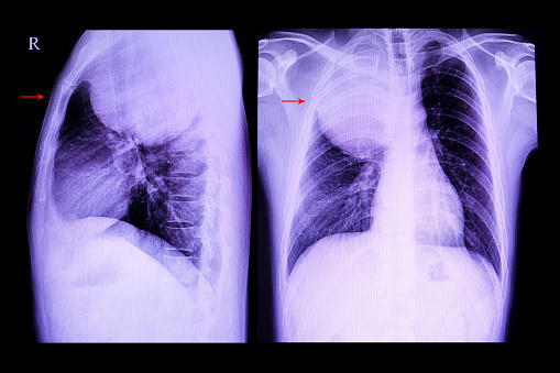
In this case, a 24-year-old woman presented with the following manifestations: dyspnea; palpitations; tachycardia; anxiety; and weight loss. Physicians evaluated her for hyperthyroidism using a combination of lab testing and imaging, and she was subsequently treated with radioiodine. Give her symptoms, was also given a computed tomography angiogram of her chest to look for pulmonary embolism.
According to the results, the patient was found to have undetectable thyroid-stimulating hormone, elevated free thyroxine (2.9 ng/dL), and elevated thyroid-stimulating immunoglobulins (399%). She was diagnosed with GD, which was confirmed by radioactive iodine uptake (RAIU) scan. The computed tomography scan of the patient’s chest revealed a large anterior mediastinal mass (7.9 × 6.9 × 6.3 cm), which was reduced in volume by 76% by radioiodine therapy.
“While the patient's thyroid labs and RAIU scan were consistent with GD, the presence of massive thymic hyperplasia was atypical,” the researchers concluded. “However, the resolution of thymic hyperplasia after radioiodine therapy, without the use of thymectomy, was similar to other reported cases.”
PubMed Link: https://pubmed.ncbi.nlm.nih.gov/32524030/
Keywords: dyspnea, palpitations, tachycardia, anxiety, weight loss, Graves’ disease, hyperthyroidism, thymic hyperplasia
Retinal Perfusion Can be Analyzed Using Optical Coherence Tomography Angiography in Patients with Inactive Graves’ Ophthalmopathy
A study which appeared in Endocrine Practice suggests the use of optical coherence tomography angiography (OCTA) can be used to analyze retinal perfusion in patients with inactive Graves’ ophthalmopathy (GO).
In this study, the research team evaluated 29 patients with inactive GO and a control group of 29 health subjects. Subsequently, they extracted and assessed vessel density (VD) data in the superficial and deep retinal OCT angiogram of the macula and the radial peripapillary capillary network (RPC). The researchers noted that OCTA was performed using RTVue XR Avanti with AngioVue, and clinical activity was analyzed using the clinical activity score, while severity was assessed using the NOSPECS classification.
Following analysis, the researchers observed that VD in the superficial OCT angiogram and in the optic nerve head (ONH) angiogram were notably lower in the GO group juxtaposed to the control group (whole en face, P=.016; parafovea, P=.026; RPC peripapillary, P=.027). The results showed no link between VD and functional parameters or the NOSPECS classification.
“Macular VD and ONH capillary density measured using OCTA were significantly lower in the study group compared to healthy controls,” the researcher authors wrote. “Noninvasive quantitative analysis of retinal perfusion using OCTA could be useful in monitoring patients with GO.”
PubMed Link: https://pubmed.ncbi.nlm.nih.gov/31859550/
Keywords: optical coherence tomography angiography, radial peripapillary capillary, Graves’ ophthalmopathy